
1.021 , 3.021, 10.333, 22.00 I ntroduc tion to Modeling and Simulation
Spring 2011
Part I – C ontinuum and partic le me thods
Applications to biophysics and bionanomechanics
Lecture 10
Markus J. Buehler
Laboratory for Atomistic and Molecular Mechanics Department of Civil and Environmental Engineering Massachusetts Institute of Technology
Content overview
I. Particle and continuum me thods
1. Atoms, molecul e s, chemistry
2. Continuum modeling approac hes and solution approaches
3. Statistical mechanics
4. Molecular dynamics, Monte Carlo
5. Visualization and data analysis
6. Mechanical proper ties – applic ation: how things fail (and how to prevent it)
7. Multi-scale modeling par adigm
8. Biological systems (simulation in biophysics) – h ow proteins work and how to model them
II. Quantum mechanical methods
1. It’s A Q uantum World: T he Theory of Quantum Mechanics
2. Quantum Mechanics: Practice Makes Perfect
3. The Many-Body Problem: Fr om Many-Body to Single- Particle
4. Quantum modeling of materials
5. From Atoms to Solids
6. Basic pr operties of mater i als
7. Advanced proper ties of materials
8. What else can we do?
Lectures 1-13
Lectures 14-26
Overview: Material covered so far…
Lecture 1: Broad introduction to IM/S
Lecture 2 : Introduction to atomistic and continuum modeling (multi-scale m odeling paradigm, difference between continuum and at omistic approac h , c a se st ud y: diff us ion)
Lecture 3 : Basic statistical mechani cs – p roperty calculation I (property calc ulat ion: microsc o pi c stat es vs. macro scopic properties, ensembles, probability density and partition function)
Lecture 4 : Property calcula tion II (Mont e Carlo, a d v a nc e d prope rty calcul ati on, introduction to chemical interactions)
Lectu r e 5: Ho w to m odel ch em i c al interactions I ( e xa mp le: mo vie o f coppe r def ormation/ di sloc ati o ns, etc.)
Lectu r e 6: Ho w to m odel ch em i c al interactions I I (EAM, a bit of ReaxFF—chemical reactions)
Lectu r e 7: Ap pli c atio n to modeling brittle materials I
Lectu r e 8: Ap pli c atio n to modeling brittle materials II
Lectu r e 9: Ap pli c atio n – A p pli catio ns to m a teri al s fai l ure
Lecture 10: Applications to biophysics and bionanomec hanics 3
Lecture 10: Applications to biophysics and bionanomechanics
Outline:
1. Protein force fields
2. Single molecule mechanics
3. Fracture of protein domains – B ell model
Goal of today’s lecture:
Force fields for organic materials, and specifically proteins
Basic introduction into modeling of biological materials
Fracture model for protein domains
1. Force fields for organic chemistry - how to model proteins
Significance of proteins
Proteins are basic building blocks of life
Define tissues, organs, cells
Provide a variety of functions and properties , su ch as mechanical stability (strength), elasticity , catalytic activity (enzyme), electrochemical properties, optica l properties, energy conversion
Molecular simulation is an important tool in the analysis of pr otein structures and protein materials
Goal here: To train you in the fundamental s of modeling techniques for proteins, to enable you to carry out protein simulations
Explain the significance of proteins ( application )
Human body: Composed of diverse array of protein materials
Eye’s cornea (collagen material)
Muscle tissue (motor proteins)
Skin (complex composite of collagen, elastin)

Cells (complex material/system based on proteins)
Imag e removed du e t o co pyrigh t restric t ion s. Human Bod y 3D View ™ i m a ge o f w h ole bo die s .
Nerve cells Blood vessels
Tendon (links bone, muscles)
Cartilage (reduce friction in joints)
Bone (structural stability)
Imag e court e sy of NIH.
Cellular structure: Protein networks

Cell nucleus
Actin network Microtubulus
(e.g. cargo)
Vimentin (extensible, flexible, provide strength)
= cyto skeleton Image co u r te s y of NI H.


Protein structures define the cellular architecture
Intermediate filaments
Imag e removed du e t o copyri gh t restri ct ion s ; see i m age now: http://ww w .nanower k.com/spot ligh t/id2 878_1. jpg . S o urc e : Fig. 2.17 in Bueh ler, Mar k us J. Atomi s ti c Mod e lin g of Materials Failure. Sp ring er, 2008.
How protein materials are made – the genetic code
Proteins: Encoded by DNA (three “letters”), utilize 20 basic building blocks (amino acids) to form polypeptides
Polypeptides arrange in complex fol ded 3D structures with specific properties
1D structure transforms into co mplex 3D folded configuration

ACGT
Four letter code “DNA”
Transcription/ translation
.. - P roline - S erine – Proline - Alanine - . .
Sequence of amino acids “polypeptide” (1D structure)

Combination of 3 DNA letters equals a amino acid
E.g. : Proline –
CCT, CCC, CCA, CCG
Folding (3D structure)
Chemical structure of peptides/proteins
Typically short
sequence

… of amino acids
side chains
Longer sequence of amino acids, often complex 3D structure
Peptide bond …
© s ourc e u n known. All right s res e rved. This con t en t is excluded fr om our Creat i ve Co mmo ns li cense . Fo r m o re inf o rma t io n , see h ttp:/ /ocw.mi t . e du/fai ruse .
R = side chain, one of the 20 natural amino acids
20 natural amino acids differ in their side chain chemistry 11
Nonpolar Amino Acids
CH 3
CH 3
CH 3
H CH 3
CH 3 CH 3 CH
CH
CH 2
CH 3
CH 2 R
CH
Forms peptide bond
+
H 3 N
C COO -
+
H 3 N
C COO -
+
H 3 N
C COO -
+
H 3 N
C COO -
+
H 3 N
C COO -
H
Glycine (Gly) G
6.0
NE
CH 2
+ -
H
Alanine (Ala) A
6.0
NE
CH 3 S
CH 2 CH 3
+ -
H
V aline (V al) V
6.0
E
CH 2
CH 2 CH 2
+ -
H
Leucine (Leu) L
6.0
E
H N
CH 2
+ -
H
Isoleucine (lle) l
6.0
E
H 3 N C COO
H
H 3 N C COO
H
H 2 N C COO
H
H 3 N C COO
H
There are 20 natural
Phenylalanine (Phe) F 5.5
E
Methionine (Met) M 5.7
E
Proline (Pro) P 6.3
NE
T ryptophan (T rp) W 5.9
E
amino acids
Polar Amino Acids (Neutral) OH
O NH 2
O NH 2
C
OH
CH 2
CH 3
HCO H
CH 2
SH
CH 2
C
CH 2
CH 2
CH 2
Difference in side
chain, R
+
H 3 N
C COO - H
+
H 3 N
C COO - H
+
H 3 N
C COO - H
+
H 3 N
C COO - H
+
H 3 N
C COO - H
+
H 3 N
C COO - H
Serine (Ser) S 5.7
NE
Threonine (Thr) T 5.6
E
T yrosine (T yr) Y 5.7
NE
Cysteine (Cys) C 5.1
NE
Asparagine (Asn) N 5.4
NE
Glutamine (Gln) Q 5.7
NE
Acidic Amino Acids
O
O - -
Basic Amino Acids
charges +
NH 2
+
O O -
C
CH 2
+ -
C CH 2
CH 2
+ -
NH 3
CH 2
HN
+ CH 2
NH CH
C NH
CH 2
CH
NH 2
H 3 N C COO
H
H 3 N C COO
H
CH 2
2
CH 2
2
CH 2
Aspartic acid (Asp) D 2.8
NE
Glutamic acid (Glu) E 3.2
NE
+
H 3 N
C COO - H
+
H 3 N
C COO -
H
+
H 3 N
C COO -
H
Histidine (His) H
7.6
E
L ysine (L ys) K
9.7
E
Ar ginine (Ar g) R
10.8
E

Image by MIT OpenCou r seWare. 12
Chemistry, structure and properties are linked
Chemical structure


Cartoon
Presence of various chemical bonds:
• C ovalent bonds (C-C, C-O, C-H, C-N..)
• Electrostatic interactions (charged amino acid side chains )
• H -bonds (e.g. between H and O)
• v dW interactions (uncharged parts of molecules)
Concept: split energy contributions
U Elec U Covalent
=0 for proteins

U total
U Metallic
U vdW
U H bond
Covalent bond described as
Ethane C 2 H 6
1. Bond stretching part (energy penalty for bond stretching)
2. Bending part (energy penal ty for bending three atoms)
3. Rotation part (energy penalty for bond rotation, N ≥ 4)
Consider ethane molec u le as “ elastic structure ”
U Covalent
U stretch
U bend
U rotate
Force fields for organics: Basic approach
=0 for proteins
U total
U Elec
U Covalent
U Metallic
U vdW
U H bond


B ond stretching
Angle Bending
B ond R otation
U Covalent
U stretch
U bend
U rot
stretch
1 k
2
2
stretch
( r r 0 )
U stretch
stretch
pairs
bend
1 k
2
2
bend (
0 )
U bend
bend
triplets
rot
1 k
2
rot
( 1 cos ( ))
U rot
rot
quadruplet s
Image by MIT OpenCou r seWare. 15
Model for covalent bonds
1 k
( ) 2
stretch

1 k
2
stretch
( r r ) 2
0
rot
1 k

2
rot
( 1 cos ( ))
bend
2 bend 0

Courte s y of the E M Bn et Ed u c ati on & Trai ni ng Commi t t ee. Use d wi th pe rmi s si o n .
16
Images cr eated for the CHARMM tutori a l by Dr. Dmit ry Kuz n e ts o v (Swi ss In sti t ute o f B i oi nformati c s ) for the EMBnet Edu c ati o n & Trai ni ng committee ( h ttp://www.embn et.org )
http://ww w.ch.embne t.org/MD_tutorial/pages/MD. P art2.html
Force fields for organics: Basic approach
=0 for proteins
U total
U Elec
U Covalent
U Metallic
U vdW
U H bond
U Elec
partial charges
q i q
j
U Elec
: Coulomb potential
( r ij )
q i q j
1 r ij


Electrostatic inter actions
vdW Inter actions
electrostatic constant
distance
q i q j
Coulomb for c es
F ( r ij ) r 2
1 ij
1 4 0 1 . 602 10 C
0
19
Image by MIT OpenCou r seWare. 17
Force fields for organics: Basic approach
U total
U Elec
U Covalent
=0 for proteins
U vdW
U Metallic
U H bond


vdW Inter actions
U vdW
Image by MIT OpenCou r seWare.
r ij
12
6
U vdW :
LJ potential
( r ij )
4
r
ij
LJ potential is particularly good m odel for vdW interactions (Argon) 18
H- bond model
=0 for proteins
U total
U Elec
U Covalent
U Metallic
U vdW
U H bond
H 2 O
D
H
DHA
H- bond
H 2 O
A
U H bond
Evaluated between acceptor (A) /donor(D) pairs
Between electronegative atom and a H- atom that is bonded to another electronegative atom
Slightly modified LJ, different parameters
R
12 R
10
U H bond :
( r
) D
5 H bon d
6 H bon d
cos 4 ( )
r ij
ij
= distance between D-A
H bond
r ij
r ij
DHA
19
Summary
=0 for proteins
U total
U Elec
U Covalent
U Metallic
U vdW
U H bond
U : Coulomb potential
( r )
q i q j
Elec
ij r
stretch
1 k
2
stretch
1 ij
2
( r r 0 )
U U U U
1 k ( ) 2
Covalent stretch
bend
rot
bend 2 bend 0
rot
1 k
2
rot
( 1 cos ( ))
12 6
r ij
U vdW :
LJ potential
( r ij )
4
r
ij
R
12 R
10
U H bond :
( r
) D
5 H bon d
6 H bon d
cos 4 ( )
ij H
bond
r ij
r ij
DHA
20

The need for atom typing
Limited transferability of potential expressions: Must use different potential for different chemistry
Different chemistry is captured in dif ferent “t ags” for atoms: Element type is expanded by additional information on particular chemical state
Tags spec ify if a C-atom is in sp 3 , sp 2 , sp or in aromatic state (that is, to capture resonance effects)
sp 3
sp 2
sp
Example atom tags : CA, C_1, C_2, C_3, C…, HN, HO, HC, …
Atom typing in CHARMM

VMD analysis of protein structure

Common empirical force fields for organics and proteins
Class I (experiment derived , simple form)
Harmonic terms;
CHARMM
pset #3
Derived from
CHARMm (Accelrys)
AMBER
OPLS/AMBER/Schrödinger
ECEPP (free energy force field)
GROMOS
Class II (more complex, derive d fro m QM )
CFF95 (Biosym/Accelrys)
MM3
UFF, DREIDING
MMFF94 (CHA R MM, Macromodel…)
vibrational
spectroscopy, gas- phase molecular structures
Very system- specific
Include anharmonic terms
Derived from QM, more general
http://ww w.ch.embne t.org/MD_tutorial/pages/MD. P art2.html
CHARMM force field
Widely used and accepted m odel for protein structures
Programs such as NAMD have implemented the CH ARMM force field
Problem set #3, nanoHUB stretchmol module, study of a pr otein domain that is part of human vimentin intermediate filaments
Application – protein folding

ACGT
Four letter code “DNA”
Transcription/ translation
.. - P roline - S erine – Proline - Alanine - . .
Sequence of amino acids “polypeptide” (1D structure)
Combination of 3 DNA letters equals a amino acid
E.g. : Proline –
CCT, CCC, CCA, CCG
Folding (3D structure)
Goal of protein folding simulations:
Predic t folded 3D structure based on poly peptide sequence
Movie: protein folding with CHARMM
de novo Folding of a Transmembrane fd Coat Protein
http://ww w.charmm- gui.org/?doc=gallery&id=23
Polypeptide chain
Images removed due to copyright restrictions.
Screenshots from protein folding video, which can be found here:
http://ww w.charmm- gui.org/?doc=gallery&id=23 .
Quality of predicted structures quite good
Confirmed by comparison of the MSD deviations of a room temperature ensemble of conformations from th e replic a-exchange simulations and experimental structures from both solid-state NMR in lipid bilayers and solution-phas e NMR on the protein in micelles)
Movies in equilibrium (temperature 300 K)

Dimer

Tetramer (increased effective bending stiffness,
interaction via overlap & head/tail domain)
Sour c e : Qi n, Z . , L . Krepl a k, and M. Bu ehl e r. “H i e rar c hi cal Struct ur e Control s Nan o mec han i c al
Pro p erti es of Vi menti n Interm edi ate Filaments.” PLoS ONE (200 9) . L i c ens e CC BY . 28
2. Single molecule mechanics
Structure and mechanics of protein, DNA, etc. molecules
Cooking spaghetti


Photo c o urtes y of Ha tM o n F lic kr .
Publ i c domai n i m age.
Photo c o urtes y of Ha tM o n F lic kr .
stiff rods cooking soft, flexible rods ( like many protein molecules )
Single molecule tensile test – “ optical tweezer”

molec u le
one end of molec u le fixed at surface
bead trapped in laser light (moves with laser)
R ep r i n t e d by p er m i s s i on fr o m M a c m illa n P u b l i s hers L t d : N a t u re .
So u r ce: T s kho v r ebo va , L . , J . T r in ick, e t a l . "Ela s t i cit y a n d Un fo l d ing o f Sin g le M o l ecu les of t h e G i a n t M u sc le Protei n Ti ti n." Na ture 387, no. 6 6 3 0 (1997 ): 308 - 1 2 . © 1 997 .
Example 1: Elasticity of tropocollagen molecules
300 nm length


14
12
10
Experimental data
Theoretical model
8
6
4
2
0
-2
0
50
100
150
200
250
300
350
Extension (nm)
The force-extension curve for stretching a single type II collagen molecule. The data were fitted to Marko-Siggia entropic elasticity model. The molecul e length and persistence length of this sample is 300 and 7.6 nm, respectively .
For ce (pN)
Entropic elasticity leads to strongly nonlinear elasticity
P h ot o co urt e sy of HatM on F lic k r .
Image by MIT OpenCou r seWare.
Courtesy of Elsevier, Inc., http://www.scien c edirect. com .
Us ed w i t h p e r m iss i on . 32
Example 2: Single protein molecule mechanics
Optical tweezers experiment
Protein structure (I27 multidomain titin in muscle)
Repr inted by p e rmi s si o n from M a c m ill an Publi s he r s L t d: N a t u r e . So urce: Tskho v rebova, L. , J . Tri n ick, e t al . "El a s t i c ity and Unfo ldin g of Single M o l e cule s of th e Gi an t Mu s cl e P r ot ein Titin . " Nat ur e 387, no. 66 30 ( 1 9 9 7 ) :
308 - 12. © 19 97.

R ep r i n t e d by p er m i s s i on fr o m M a c m illa n P u b l i s hers L t d : N a t u re .
So u r ce: M a r s za lek, P . , H . L u , e t a l . "M ech a n i c a l Un fo ld i n g I n t e rme d ia t e s in T i t i n M o du les . " Na ture 402, no. 6 75 7 (1999 ): 100 -3 . © 1 999 .
http://ww w.nature.com/nature /journal/v387/n6630 /pdf/387308a0.pdf http://ww w.nature.com/nature /journal/v402/n6757 /pdf/402100a0.pdf 33
Example 3: Single DNA molecule mechanics
plateau regime (break ing of bonds)


Courte sy of El se vie r, Inc., h t t p ://www. scien c e dir ec t . c o m . Used wi th permission.
Plots of stretching force against relative extension of the single DNA molecule (experimental results)
Structural makeup of protein materials
Although very diverse , all protein materials have universal “protocols” o f how they are made
How protein materials are made–the genetic code
Proteins: Encoded by DNA (three “letters”), utilize 20 basic building blocks (amino acids) to form polypeptides
Polypeptides arrange in complex fol ded 3D structures with specific properties

1D structure transforms into co mplex 3D folded configuration
ACGT
Four letter code “DNA”
Combination of 3 DNA letters (=codon) defines one amino acid
E.g. : Proline –
CCT, CCC, CCA, or CCG
Transcription/ translation
.. - P roline - S erine – Proline - Alanine - . .
Sequence of amino acids “polypeptide” (1D structure)
Folding (3D structure)
36
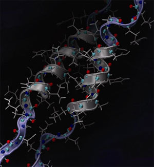
Alpha-helix (abbreviated as AH)
Concept: hydrogen bonding (H-bonding)


e.g. between O and H in H 2 O Bet w een N and O in proteins


Drives formation of helical structures AHs found in: hair, cells, wool, skin, e t c.
A dapted f r om Bal l , D., Hil l , J., et al. T h e Basi c s of Ge n e ra l, Org ani c, and Bi o l og ic al Ch e m is t r y. Fla t worl d Knowl edge, 2011. C o urtesy of Flatworld Kn owl e dge.
Sour c e : Qi n, Z . , L . Krepl a k, and M. Bu ehl e r. “Hi e rarc hi cal str u ctu re co n trol s nan o mec ha ni c al pr operti es of vi menti n in t e r m ed ia t e fi la me n t s . ” PL o S O N E (2009 ) . Li c e n s e C C BY.
Primary, secondary, tertiary structure


A dapted f r om Bal l , D., Hil l , J., and R. S c ott. T h e Ba sics o f G e ne ra l , O r g a nic , a n d Bi o l og ic al Ch e m is t r y . Fl atworl d Kn owl e dg e, 20 1 1 . Courte s y o f Fl atworl d Kn owl e dge.
38
Beta-sheets (abbreviated as BS)
Beta-sheet
Images r e mo ve d d u e to c o py ri ght restri cti o n s .
Found in many mechanic ally relevant proteins
Spider silk Fibronectin
Titin (muscle tissue)
Amy loids (Alz heimer’s disease) 39
Amyloid proteins (Alzheimer’s disease)
Pl ease s e e Fig. 8 from htt p :/ /w eb.mi t .edu /m bu e h l e r/www / p aper s /fi nal _ JCTN _ p r e pri n t.pdf .
3. Fracture of protein domains – Bell model
How to apply load to a molecule
(in molecular dynamics simulations)
Steered molecular dynamics (SMD)
Steered molecular dynamics used to apply forces to protein structures
v
Virtual atom
moves w/ velocity v k
x


end point of molec u le
Steered molecular dynamics (SMD)
Steered molecular dynamics used to apply forces to protein structures
v


Virtual atom f
moves w/ velocity v k
x
x
f k ( v t x )
v t
end
SMD spring constant
point of
molec u le
f k ( v t x )
SMD
deformation speed vector
time
Distance between end point of molecule and virtual atom
k
x
v
k
x
SMD mimics AFM single molecule experiments
Atomic force microscope
v
f
x
SMD is a useful approach to probe the nanomechanics of proteins (elastic deformation, “plastic” – permanent deformation, etc.)
Example: titin unfolding (CHARMM force field)
Unfolding of titin molecule
X : breaking
Force (pN)
X
X
Titin I27 domain: Very resistant to unfolding due to parallel H-bonded strands


Displacement (A)
47
Keten and Buehler, 2007
Protein unfolding - R eaxFF

F
AHs
PnIB 1AKG
F


ReaxFF modeling
Buehl e r, M. " Hi erar c h i c al Chemo - nan o mecha n i c s of Protei n s : E n tropi c El a s ti ci ty, Protei n Unfol d i n g
a n d Mo lecu lar F r a c t u re . " J o urnal of M e chani c s and Mate rial s and S t ruc t ures 2 , no. 6 ( 2 007). 48
Protein unfolding - CHARMM

Covalent bonds don’t break
CHARMM modeling
Comparison – CHARMM vs. ReaxFF


Application to alpha-helical proteins
51

Vimentin intermediate filaments
Sour c e : Qi n, Z . , L . Krepl a k, et al. "Hi e rarc hi cal Stru ctu re Control s Nan o mec han i c al Properti es of Vi menti n Intermedi a te F i laments." PLoS ON E 4, no. 1 0 ( 2009) .
d o i : 10 .1 371/journal.p o ne .000 7294 .
Li c e n s e C C BY. 52
Cells
Vimentin intermediate filament
Filaments
Protein molecule
Chemical bonding
53
Sour c e : Qi n, Z . , L . Krepl a k, et al. "H i e rarc hi cal Structu r e Control s N a no mech a n ic a l P r o p er t i es o f Vime n t in I n t e r m e d ia t e F i la me n t s . " PLoS ONE (2009 ) . L i c ense C C B Y .


hair, hoof
Intermediate filaments – occurrence

neuron cells (brain)


fibroblast cells (make collagen)
cell nucleus
I m a g e o f n e ur on a n d c e ll nuc leus © s ou rces unkn ow n . All r ig h t s r e served . T h i s content is e x c l ud ed from our Creative Common s lice nse . F o r m o re in fo r m a t io n , se e h t t p ://ocw.m i t .e d u / f a i r u se .
Alpha-helical protein: stretching


ReaxFF modeling of AH stretching
M. Buehler, JoMMS, 2007
A: Firs t H-bonds break (turns open) B: Stretch covalent backbone
C: Backbone breaks 55
What about varying pulling speeds?

12,000
1,500
1,000
500
8,000
0 0
0.2
0.4
4,000
0 0
50
100
150
200
Strain (%)
v = 65 m/s v = 45 m/s v = 25 m/s v = 7.5 m/s v = 1 m/s model
model 0.1 nm/s
Force (pN)
Variation of pulling speed
Image by MIT OpenCou r s e Ware. After Ack b arow an d Buehl e r, 2007.
Force at AP (pN)
Force at angular point f AP =fracture force
f AP ~ ln v

Pulling speed (m /s)
General results…
Rupture force vs. pulling speed

f AP
R ep r i n t e d by p er m i s s i on fr o m M a c m illa n P u b l i s hers L t d : N a t u re M a t e r i a l s .
Sour c e : Buehl e r, M., and Y. Yung. " C hemom e c h ani c al Behavi ou r of Protei n C o n s ti tuent s ." Nature Mater ial s 8, no. 3 (2 0 0 9 ) : 175 -88 . © 2 009 .
How to make sense of these results?
A few fundamental properties of bonds
Bonds have a “ bond energy ” ( energy barrier to break)
Arrhenius relationship gives probability for energy barrier to be overcome, given a temperature
E b
p exp
k B T

All bonds vibrate at frequency
Bell model
Probability for bond rupture (Arrhenius relation)

E b
p exp
k B T
Boltzmann constant
temperature
distance
height
Bell model
Probability for bond rupture (Arrhenius relation)
f f AP
p exp
E b f x B
k B T
force applied ( lower energy barrier )
Boltzmann constant
temperature
distance
height
Bell model
Probability for bond rupture (Arrhenius relation)
p exp
E b f x B
k B T
0
Off-rate = probability times vibrational frequency
( E b f x b
exp ) 1
0 k T
b
0 p
1 1 0 13 1 / sec
Bell model
Probability for bond rupture (Arrhenius relation)
p exp
E b f x B
k B T
Off-rate = probability times vibrational frequency
1
( E b f x b ) 13
0 p
0
exp
k b T
0 1 10
1 / sec
“How often bond breaks per unit time”
Bell model
Probability for bond rupture (Arrhenius relation)
p exp
E b f x B
k B T
Off-rate = probability times vibrational frequency
( E b f x b ) 1 13
0 p
0
exp
k b T
0 1 10
1 / sec
bond lifetime (inverse of off-rate)

Bell model
t ???
x
x
x / t v
t
x / t v
pulling speed (at end of molecule)
Bell model
t
x x
broken turn
x / t v
x


x t
x / t v
pulling speed (at end of molecule)
Structure-energy landscape link
x b


x x b 1
t
( E b
f x b )
0
exp
k b T
Bell model
x / t v
x
broken turn
t
x x b t
Bond breaking at
x b (lateral applied displacement):
( E b f x b )
x b
0
exp
k b T
x b
x / t v
1 /
pulling speed
Bell model
( E b f x b )
0 exp
k b T
x b v
Solve this expression for f :
Bell model
( E b f x b )
0 exp
k b T
x b v
Solve this expression for f :
( E b
f x b )
ln(
x )
ln v
ln(..)
0 b
k b T
E b f x b k b T ln v ln( 0
x b )
E b k b
T ln v
ln( 0
x b )
k b T
k b T E b
f
x b
ln v
x b
x b k b T
ln( 0
x b )
k b T
k b T
E b
f ln v
x b
ln( 0
x b
x b )
k b T
k b T
k b T
E b
f ln v
x b
ln 0
x b
x b
exp
k b T 73
Simplification and grouping of variables
Only system parameters, [distance/length]
k b T
k b T
E b
f ( v ; x b , E b )
ln v
x b
ln 0
x b
x b
exp
k b T
: v 0
0
x b
exp
E b
k b T
Bell model
( E b f x b )
0 exp
k b T
x b v
Results in:
f ( v ; x , E
) k b T
ln v
k b T
ln v
a ln v b
x
x
b b 0
b b
a k B T
x b
x
b k B T
ln v
0
b
f ~ ln v behavior of strength

f ( v ; x b , E b ) a ln v b
Force at AP (pN)
Pulling speed (m /s)
E b = 5.6 kcal/mol and x b = 0.17 Ǻ (results obtained from fitting to the simulation data)
f ( v ; x b , E b ) a ln v b
E b
Force at AP (pN)
Scaling with E b : shifts curve
k B T
k B T
Pulling speed (m /s)
E b
a b
x b x b
ln v 0
v 0 0
x b
exp
k b T
7 7
f ( v ; x b , E b ) a ln v b
x b
Force at AP (pN)
Scaling with x b : changes slope
k B T
k B T
Pulling speed (m /s)
E b
a b
x b x b
ln v 0
v 0 0
x b
exp
k b T
7 8
Simulation results


Courtesy of IOP Publishing, Inc. Used with permission. Source: Fig. 3 from Bertaud, J., Hester, J. et al. "Energy Landscape, Structure and
Rate Effects on Strength Properties of Alpha-helical Proteins." J Phys.: Condens. Matter 22 (2010): 035102. doi:10.1088/0953-8984/22/3/035102.

Mechanisms associated with protein fracture


Change in fracture mechanism
Single AH structure
FDM : Sequential HB break ing
SDM : Concurrent HB break ing
(3..5 HBs)
Simulation span: 250 ns
Reaches deformation speed O(cm/sec)
Courtes y of Nati onal Academ y of Sci e n c e s , U. S. A. Use d w i th permi s si o n . Sour c e : Ackbarow, Theo dor, et al. " H i e rar c hi es, Mul t i p l e Ener gy Barri er s, and Ro bu stn e s s Go v e r n t h e Fractu r e Mec h ani c s of Al pha- hel i cal and Beta- s h eet Protei n D o mai n s. " PN A S 104 ( O c t obe r 1 6 , 20 0 7 ) : 1 6 4 1 0 - 5. Copy ri g h t
200 7 National Acad e m y of Scie nces, U. S . A. 81
Analysis of energy landscape parameters


Energy single H-bond: ≈ 3-4 kcal/mol
What does this m e an???
82
Courtes y of Nati onal Academ y of Sci e n c e s , U. S. A. Use d w i th permi s si o n . Sour c e : Ackbarow, Theo dor, et al. " H i e rar c hi es, Mul t i p l e Ener gy Barri er s, and Ro bu stn e s s Go v e r n t h e Fractu r e Mec h ani c s of Al pha- hel i cal and Beta- s h eet Protei n D o mai n s. " PN A S 104 (O ctober 16, 2007): 16410-5. Copy right 200 7 National Acad e m y of Scie nces, U. S . A.
H- bond rupture dynamics: mechanism
I: All HBs are intact
Courtes y of Nati onal Academ y of Sci e n c e s , U. S. A. Use d w i th permi s si o n . Sour c e : Ackbarow, Theo dor, et al. " H i e rar c hi es, Mul t i p l e Ener gy Barri er s, and Ro bu stn e s s Go v e r n t h e Fractu r e Mec h ani c s of Al pha- hel i cal and Beta- s h eet Protei n D o mai n s. " PN A S 104 ( O c t obe r 1 6 , 20 0 7 ) : 1 6 4 1 0 - 1 5 .

C o py ri g h t 20 0 7 Nati onal Aca d em y of S c i e n c e s , U.S.A.
II: Rupture of 3 HBs – s imultaneous ly ; within ≈ 20 ps
III: Rest of the AH relaxes – s lower deformation…